March 25, 2026


The Latest Cardiac and Vascular News
March 25, 2026
Members of the SHERPA project team
Members of the SHERPA project team
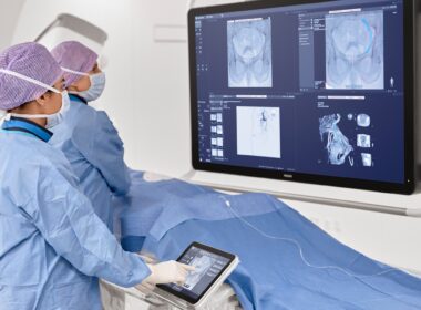
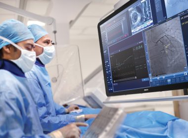
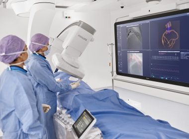
Philips Azurion interventional oncology procedure
Philips Azurion interventional oncology procedure
SHERPA project logo
SHERPA project logo
March 3, 2026 With a total budget of EUR 21.5 million, the four-year SHERPA research project is coordinated by Philips and co-funded by the EU Innovative Health Initiative (IHI) and industry partnersThe project focuses on developing automated workflows for minimally invasive neurovascular and tumor treatments – clinical procedures where staffing levels and specialized expertise are in short supplyThe clinical studies are being conducted at University Medical Center Utrecht, St Antonius Hospital, University Medical Center Hamburg-Eppendorf, Hôpital Bicêtre AP-HP, and Hospital de la Santa Creu i Sant PauOn March 4 (16:30 – 17:30 CET), SHERPA will be featured at the European Congress of Radiology 2026 in a dedicated session Amsterdam, the Netherlands – Royal Philips (NYSE: PHG, AEX: PHIA), a global leader in health technology, today announced that the Philips-coordinated SHERPA research consortium has initiated seven clinical studies to demonstrate the benefits of AI- and robotics-assisted workflows in minimally invasive treatments for brain aneurysms* and liver tumors. Staff shortages, coupled with the complex nature of the work, place significant pressure on interventional radiologists and interventional neuroradiologists who perform these procedures. The SHERPA consortium aims to validate AI-powered technologies in imaging, data visualization, procedure planning and guidance, clinical decision support, and patient pathway orchestration. These technologies are designed to automate repetitive, time-consuming tasks, support decision-making, and accelerate learning to ease the workload of interventional (neuro)radiologists. The four-year research project is co-funded by the industry partners through in-kind contributions and additional resources, as well as by the European Union Innovative Health Initiative (IHI), and comprises 16 partners from seven European countries. “Over the past year, the SHERPA project has brought together a talented team of researchers who have developed a technology framework designed to make workflow automation a reliable companion – a ‘sherpa’ – for interventional (neuro)radiologists as they navigate some of their most complex procedures,” said Bert van Meurs, Chief Business Leader, Image Guided Therapy, at Royal Philips. “By providing a template for the future adoption of AI-enabled assistive technologies and accelerating the associated learning curve, this framework will help address the increased demand for interventional (neuro)radiologists in Europe and beyond.” “Demand for image-guided, minimally invasive procedures has outpaced the growth of the specialized workforce,” said Irene van der Schaaf, Professor Neurovascular Interventional Radiology, UMC Utrecht. “At the same time, procedures are becoming more complex and technology is advancing rapidly, making it harder for teams to keep pace. In interventional radiology, technology and human touch go hand in hand. Physicians must manage highly technical tasks – sometimes away from the patient – while continuing to provide personal care and reassurance. The SHERPA project aims to develop AI-enabled tools that simplify routine tasks, enhance patient care, and support the training of the next generation of interventional (neuro)radiologists.” On March 4, 2026, from 16:30 to 17:30 CET, SHERPA will be featured at the European Congress of Radiology (ECR) 2026 in a dedicated session titled: “Assistive Technologies for Interventional Radiologists: Enhancing Decision-Making and Streamlining Workflow with Innovations and AI-Powered Solutions.” Click here to view the agenda and register to attend the session online. Innovation through public-private partnershipThe SHERPA public-private partnership consortium comprises five medical technology industry partners, five academic partners, and five research organization and medical association partners: Industry partners: Philips, Medtronic, Sim&Cure, Interventional Systems, Barco.Academic partners: University Medical Center Utrecht (The Netherlands), University Medical Center Hamburg-Eppendorf (Germany), Hôpital Bicêtre AP-HP (France), Hospital de la Santa Creu i Sant Pau (Spain), St Antonius Hospital (The Netherlands).Research organization and medical association partners: European Institute for Biomedical Imaging Research – EIBIR, Cardiovascular and Interventional Radiological Society of Europe – CIRSE, European Society of Minimally Invasive Neurological Therapy – ESMINT, Eindhoven University of Technology – TU/e (The Netherlands), Human-Factors-Consult – HFC (Germany). Addressing staff shortages and patient access by relieving the pressure on physiciansThe World Health Organization (WHO) predicts a shortage of 600 thousand physicians in the European Union (EU) by 2030 [1]. Some of the most acute physician shortages are in interventional radiology, even in countries such as the UK that have advanced healthcare systems [2]. Among the many different procedures conducted by interventional (neuro)radiologists, treatments of brain aneurysms and liver tumors are some of the most complex and time consuming. Both rely on CT and/or MR imaging for diagnosis, real-time imaging for procedure guidance, and a high level of precision in the placement of therapeutic devices such as blood-clot inducing platinum coils to seal off a brain aneurysm or the percutaneous insertion of ablation needles to treat a liver tumor. As a result, these procedures require a very high level of operator expertise and training, which can limit patient access. The SHERPA project aims to provide interventionists with AI-powered assistive technologies that automate repetitive tasks and support decision-making across the entire workflow. By doing so, they will accelerate learning curves and improve precision and safety in complex minimally invasive interventions such as brain aneurysm repair and liver tumor ablation. During the first year of the project, the consortium successfully developed AI algorithms to help identify brain aneurysms that need treatment and algorithms to optimize patient selection and therapy planning for liver tumor ablation. They also developed robotic technology to improve procedure precision and reduce difficulty, and AI software to confirm treatment success. These have now been integrated into orchestrated end-to-end workflows for both types of procedure. SHERPA project clinical studiesOver the next three years of the four-year SHERPA project, the consortium partners will conduct a series of clinical studies to refine these assistive technologies and assess the benefits in terms of the patient experience, workload optimization, interventionist satisfaction, and performance. The five brain aneurysm studies will focus on AI-driven aneurysm detection, risk prediction, and precise treatment planning: RADAR: AI-based aneurysm detection based on CT and MR imaging.Aneurysm@risk: AI-based algorithm to predict aneurysm growth and rupture risk.ASSIST: AI-supported device selection and positioning guidance.INTERACT: Automatic collimation and projection angle suggestions to optimize imaging for procedural guidance.SAFO: Evaluation of a digital remote follow-up solution for brain aneurysm patients, enabling standardized monitoring, enhanced coordination, and seamless care across the patient pathway. The liver and lung tumor studies will leverage advanced imaging and robotic-assisted biopsy technologies, respectively, to drive greater diagnostic precision and procedural efficiency. MISTRAL: Evaluation of new Cone Beam CT workflows to optimize imaging for percutaneous liver ablations.RHODES: Evaluation of robotic-assisted versus free-hand lung biopsies with a focus on operability and device efficiency. Together, these studies will generate insights to support the acceptance and adoption of AI-based smart assistive technologies by the wider interventional radiology community. The SHERPA website can be viewed at: https://sherpa-ihi.eu. Watch videos and interviews about SHERPA here. The CORDIS project page for SHERPA can be accessed here, and the IHI factsheet for SHERPA can be found here. Notes and references* A brain aneurysm is a weak, bulging spot in a blood vessel in the brain, which can leak or rupture.[1] Zapata T, Muscat NA, Falkenbach M, and Wismar M. WHO Report – From Great Attrition to Great Attraction: Countering the Great Resignation of Health and Care Workers. Eurohealth, Vol.29, No.1, 2023. https://iris.who.int/server/api/core/bitstreams/488b01ab-a066-4558-a345-476570fe2802/content[2] The Royal College of Radiologists: Clinical radiology – UK workforce census 2020 report. https://www.rcr.ac.uk/media/3gjdr23o/clinical_radiology_census_report_2020.pdf This project is supported by the Innovative Health Initiative Joint Undertaking (IHI JU) under grant agreement No 101194744. The JU receives support from the European Union’s Horizon Europe research and innovation program and life science industries represented by COCIR, EFPIA, Europa Bío, MedTech Europe and Vaccines Europe. SHERPA is Funded by the European Union. Views and opinions expressed are however those of the author(s) only and do not necessarily reflect those of the European Union or Innovative Health Initiative Joint Undertaking. Neither the European Union nor the granting authority can be held responsible for them. Media ContactSteve KlinkPhilips Medical and Scientific CommunicationsTel: +31 6 10888824E-mail: steve.klink@philips.com Joost MalthaPhilips Global External RelationsTel: +31 6 10558116E-mail: joost.maltha@philips.com About Royal PhilipsRoyal Philips (NYSE: PHG, AEX: PHIA) is a leading health technology company focused on improving people’s health and well-being through meaningful innovation. Philips’ patient- and people-centric innovation leverages advanced technology and deep clinical and consumer insights to deliver personal health solutions for consumers and professional health solutions for healthcare providers and their patients in the hospital and the home.Headquartered in the Netherlands, the company is a leader in diagnostic imaging, ultrasound, image-guided therapy, monitoring and enterprise informatics, as well as in personal health. Philips generated 2025 sales of EUR 18 billion and employs approximately 64,800 employees with sales and services in more than 100 countries. News about Philips can be found at www.philips.com/newscenter.
Attachments
Members of the SHERPA project team
Philips Azurion interventional oncology procedure
SHERPA project logo

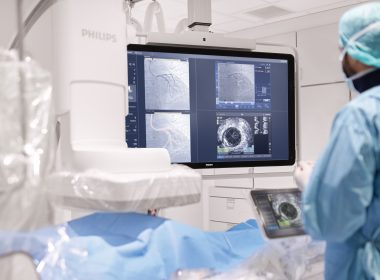
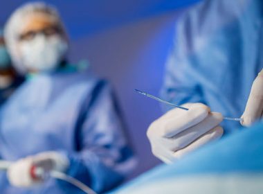
Interventional oncology procedure on Philips Azurion
Interventional oncology procedure on Philips Azurion
Members of the PreciseOnco consortium team
Members of the PreciseOnco consortium team
PreciseOnco research consortium logo
PreciseOnco research consortium logo
February 3, 2026 Co-funded by the EU’s Innovative Health Initiative (IHI), the Philips-coordinated research consortium will combine advanced medical imaging, robotic guidance technologies, and AI to standardize and enhance the precision of minimally invasive cancer treatmentsFive-year research program with total budget of EUR 23.9 million includes five clinical studies to validate the technical solutions, with the aim of setting a new benchmark for precision, safety, and efficiency in minimally invasive cancer care Amsterdam, the Netherlands – Royal Philips (NYSE: PHG, AEX: PHIA), a global leader in health technology, today announced that the PreciseOnco consortium, coordinated by Philips, has been awarded funding from the EU’s Innovative Health Initiative (IHI) to advance precision cancer treatment through the integration of advanced medical imaging, robotic assistance, and minimally invasive therapies. The EUR 14.9 million public funding will be complemented by EUR 9 million in in-kind contributions and additional resources from industry partners, supporting a five-year research and innovation program that also includes five clinical studies. “Smart imaging and robotic precision are opening new possibilities for minimally invasive cancer treatment,” said Bert van Meurs, Chief Business Leader for Image Guided Therapy at Royal Philips. “By combining spectral imaging, AI-powered software and robotic guidance, we’re developing new solutions to help physicians treat cancer with greater precision, confidence and speed. This leads to better outcomes for patients and a more efficient use of hospital resources. The PreciseOnco partnership brings together technology, clinical expertise and patient perspectives to help improve the way cancer care is delivered.” Innovation through public-private partnershipThe PreciseOnco public-private partnership unites experts from industry, research organizations, medical societies and leading European hospitals: Industry partners: Philips, Quantum Surgical, and IGEAResearch organization: European Institute for Biomedical Imaging Research (EIBIR)Medical society: Cardiovascular and Interventional Radiological Society of Europe (CIRSE)Academic partners: University Hospital Cologne (Uniklinik Koeln) in Germany, University Medical Center Utrecht (UMCU) and Leiden University Medical Center (LUMC) in the Netherlands, and two major university hospital networks in France: Assistance Publique–Hôpitaux de Paris (APHP; Hôpital Henri-Mondor) and Hospices Civils de Lyon (HCL) By uniting Europe’s leading clinical expertise and industry innovation, PreciseOnco aims to set a new benchmark for precision, safety, and efficiency in minimally invasive cancer care. The rising cancer burden and precision treatment needsCancer remains a leading health challenge, with approximately 2.7 million new diagnoses in Europe each year [1] with projections suggesting over 35 million new cancer cases globally by 2050, representing a 77% increase from 2022 [2]. In response to this growing challenge, interventional oncology has emerged as a rapidly evolving discipline that integrates medical imaging, oncology, and minimally invasive treatment approaches [3]. Such treatments – including targeted ablation, radioembolization, and electrochemotherapy – offer advantages over traditional surgery, such as shorter recovery times and fewer side effects; however, their effectiveness strongly depends on the performance of medical imaging and the precision with which physicians can guide instruments to tumors. Bringing advanced technology into the interventional roomThe PreciseOnco consortium aims to develop a suite of integrated technologies designed to take the next leap in precision cancer care. Central to the research and innovation program is spectral imaging, an advanced form of CT and cone-beam CT that captures significantly richer information about tissue composition than conventional imaging, enabling more confident differentiation between tumors, vasculature, and healthy tissue. By analyzing X-rays at different energy levels, spectral imaging enables physicians with greater clarity to see exactly what tissues they are treating. To complement this advanced imaging technology, PreciseOnco will integrate robotic guidance systems that use real-time imaging data to guide interventional instruments (one or multiple needles) with sub-millimeter precision. The consortium will also look to advance electrochemotherapy, a minimally invasive technique that combines locally administered electrical pulses with chemotherapy to selectively treat cancer tissue, with the aim of maximizing tumor control while preserving surrounding healthy tissue. Crucially, all these technologies will be powered by AI algorithms designed to enhance image quality, reduce radiation dose, streamline advanced visualization software and provide real-time intra-procedural feedback on treatment success. This would allow physicians to confirm that tumors have been fully treated before the patient leaves the operating room. Clinical studiesThe project is structured into multiple work packages covering spectral imaging technology development, AI based image processing, robotic guidance integration, multi-center clinical validation, and health economic assessment. PreciseOnco will conduct five clinical studies spanning multiple cancer types and interventional workflows, ensuring robust validation in real world clinical settings: VISTA (Virtual Spectral Imaging for Superior Thermal Ablation Guidance): Evaluating spectral imaging to improve liver, kidney ablation procedures and liver radioembolization.SPOT ON (Spectral angiography-computed tomography to Optimize percutaneous Tumor ablation): Assessing spectral CT for better tumor targeting and treatment planning.HORA EST HCC 2: Combining thermal ablation with transarterial chemoembolization (TACE) in a single session for improved hepatocellular carcinoma treatment.SPECTRA-L (Spectral Performance Evaluation of a CT-Equipped Therapeutic Radiology Angio Suite in Liver): Testing spectral imaging for transarterial chemoembolization (TACE) procedures.LASER (Locoregional therApies Spectral Evaluation of Responses): Developing imaging biomarkers to predict treatment success across multiple cancer types and interventional techniques. Together, these studies will generate evidence to support the adoption of spectral imaging and robotic guidance across European cancer centers, extending access to advanced minimally invasive treatments to a larger patient population. Philips and innovation public-private partnershipsPublic-private partnerships play a vital role in advancing healthcare innovation. With over 40 years of experience in shaping and leading such partnerships, Philips sees them as a powerful complement to its global R&D programs, helping build sustainable ecosystems that foster innovation. Philips’ broad imaging, image-guided therapy, and healthcare informatics portfolio helps clinicians visualize, assess, guide treatment, and confirm outcomes – supporting more precise and personalized care. With innovations like Verida detector-based spectral CT fully powered by AI and Azurion image-guided therapy system, Philips continues to push the boundaries of imaging technology, advanced visualization and minimally invasive therapy worldwide. The PreciseOnco project website will be available soon at www.preciseonco.eu. The CORDIS project page for PreciseOnco can be accessed here. The IHI factsheet for PreciseOnco can be found here. References[1] European Cancer Information System (ECIS) – jrc_CancerEstimates2022_factsheet.pdf.[2] World Health Organization (WHO) – Global cancer burden growing, amidst mounting need for services.[3] Global Interventional Oncology Market report – Interventional Oncology Market Size and Growth Analysis Report. This project is supported by the Innovative Health Initiative Joint Undertaking (IHI JU) under grant agreement No. 101252582. The JU receives support from the European Union’s Horizon Europe research and innovation program and life science industries represented by COCIR, EFPIA, EuropaBio, MedTech Europe and Vaccines Europe. PreciseOnco is funded by the European Union, private members, and those contributing partners of the IHI JU. Views and opinions expressed are, however, those of the author(s) only and do not necessarily reflect those of the aforementioned parties. Neither of the aforementioned parties can be held responsible for them. For further information, please contact: Joost MalthaPhilips Global External RelationsTel.: +31 610 558 116E-mail: joost.maltha@philips.com Steve KlinkPhilips Medical and Scientific CommunicationsTel.: +31 610 888 824E-mail: steve.klink@philips.com About Royal Philips Royal Philips (NYSE: PHG, AEX: PHIA) is a leading health technology company focused on improving people’s health and well-being through meaningful innovation. Philips’ patient- and people-centric innovation leverages advanced technology and deep clinical and consumer insights to deliver personal health solutions for consumers and professional health solutions for healthcare providers and their patients in the hospital and the home.Headquartered in the Netherlands, the company is a leader in diagnostic imaging, ultrasound, image-guided therapy, monitoring and enterprise informatics, as well as in personal health. Philips generated 2024 sales of EUR 18 billion and employs approximately 67,800 employees with sales and services in more than 100 countries. News about Philips can be found at www.philips.com/newscenter.
Attachments
Interventional oncology procedure on Philips Azurion
Members of the PreciseOnco consortium team
PreciseOnco research consortium logo
December 15, 2025

November 19, 2025Amsterdam, the Netherlands – Royal Philips (NYSE: PHG, AEX: PHIA), a global leader in health technology, today announced an extended partnership with Cortechs.ai, a pioneer in quantitative neuroimaging solutions. Together, the companies will integrate Cortechs.ai’s advanced AI-enabled neuroimaging analytics directly into Philips’ MR systems [1], empowering clinicians with faster, more objective, and reproducible insights into brain health. The collaboration strengthens Philips’ leadership in precision neuro diagnostics, combining Philips’ next-generation MR technologies with Cortechs.ai’s AI-driven quantitative neuro imaging post processing software [1] to transform how neurological diseases are detected, monitored, and managed.Combining complementary expertise for smarter brain imagingUp to 25% [2] of all MR procedures are brain scans, and radiology departments face growing demand amid increasing staff shortages. At the same time, the number of patients with neurological conditions such as Alzheimer’s disease, multiple sclerosis (MS), and brain tumors continue to rise globally. Current MR interpretation of neurodegenerative diseases or neuro oncology cases depends heavily on visual assessment, introducing variability and requiring significant expertise and time in an already overloaded system.Through this extended partnership, Philips and Cortechs.ai aim to deliver more standardized and data-driven imaging. By integrating quantitative analysis tools such as brain volumetrics, lesion quantification, and tumor tracking directly into the Philips MR imaging workflow [1], clinicians gain objective, numerical data alongside images – enabling faster, more consistent, and confident diagnoses.Empowering clinicians with precision and speedThe integration of Cortechs.ai’s NeuroQuant® solutions for Alzheimer’s, MS and brain tumors within Philips’ Smart Reading environment – a cloud-based AI platform that combines imaging, reading, and reporting directly on the MR systems [1] – marks a major step toward precision neuroimaging.With this integration, clinicians can automatically receive AI-generated quantitative reports within their standard MR workflow on the MR system or PACS, without adding extra steps for technologists. The zero-click process ensures quality-checked data and standardized outputs, helping radiologists interpret brain scans more objectively, faster and with greater confidence.“By extending our partnership with Cortechs.ai, we are accelerating the transition to fully quantitative, AI-powered neuroimaging,” said Ioannis Panagiotelis, PhD, Business Leader, MRI, Philips. “This integration brings precision, reproducibility, and efficiency to neurological MR, helping clinicians make faster, more confident decisions for patients living with complex brain conditions.”Driving productivity and improving care through AIPhilips is driving productivity and improving care through deep AI integration that tackles some of healthcare’s most pressing challenges – from workforce shortages to the growing demand for advanced diagnostics. By automatically generating quantitative reports within existing workflows, AI helps radiologists save time, improve consistency, and boost throughput. Objective numerical biomarkers enhance diagnostic accuracy, enabling more reproducible assessments and confident comparisons across time and sites. And by minimizing manual post-processing through automated, quality-controlled workflows, technologists can focus more on what matters most – delivering a better, more personalized patient experience.“Cortechs.ai’s mission has always been to empower clinicians with actionable, quantitative insights,” said Kyle Frye, Chief Executive Officer, Cortechs.ai. “By combining our advanced neuro analytics with Philips’ leading MR platform and AI infrastructure, we’re making it easier to personalize brain care and track neurological changes with precision.”Setting a new standard in precision neuro diagnosticsCortechs.ai’s NeuroQuant® solutions are already available on Philips Advanced Visualization Workspace. With this extended partnership, Philips reinforces its commitment to advancing the field of neurology – empowering healthcare providers to deliver better care for more people, in less time, with greater confidence.
November 17, 2025Innovation uses AI to track and visualize tiny repair devices* through the beating heart, helping clinicians navigate in 3D with enhanced clarity and confidence**Amsterdam, the Netherlands – Royal Philips (NYSE: PHG, AEX: PHIA), a global leader in health technology, today introduced DeviceGuide, an AI-powered device tracking* solution that assists physicians during one of interventional cardiology’s most technically demanding procedures: repairing leaking heart valves through a minimally invasive approach. Built on Philips’ EchoNavigator platform, this software brings AI directly into the procedure room, translating complex imaging into intuitive, real-time visual guidance that helps clinicians navigate the beating heart with greater clarity and confidence. It will be previewed at London Valves 2025 (November 16-18), one of the world’s leading meetings for structural heart specialists.“With DeviceGuide, we’re bringing AI into the heart of the procedure room, and into the heart itself,” said Dr. Atul Gupta, Chief Medical Officer, Diagnosis & Treatment at Philips. “This is Philips’ first AI assisting physicians in real time to visualize and guide heart valve treatment devices* as they navigate the beating heart. It’s helping doctors in the moment as they are helping their patients with structural heart disease.”Minimally invasive treatment of the mitral valveA leaking heart valve, known as mitral valve regurgitation, is a condition in which blood leaks backward through the heart’s mitral valve. It affects more than 35 million [1] adults worldwide, leaving many short of breath, fatigued, and struggling with everyday activities like climbing stairs or walking short distances. Many also live with anxiety or fear, knowing their heart isn’t pumping efficiently. If left untreated, severe cases can lead to heart failure and other serious complications.For patients who are too frail for open-heart surgery, minimally invasive transcatheter repair techniques such as mitral transcatheter edge-to-edge repair (M-TEER) offer a vital treatment option. During these procedures, physicians repair a leaking valve through a tiny incision in the top of the leg in the groin area, guiding long, flexible instruments through the blood veins and navigating a miniature repair device into the beating heart.Clinicians must view and interpret both X-ray and ultrasound images on multiple screens, coordinate movements between two operators, and make precise adjustments to grasp the moving valve leaflets, position the repair device, and confirm the result in real time. The process demands accuracy, coordination, and experience from the team. This is where DeviceGuide can help with its 3D navigation support.How AI is helpingDeviceGuide uses an AI algorithm to automatically track the tiny repair device as it moves through the beating heart, intelligently combining live echo and X-ray images. It creates a virtual 3D model of the treatment device in real time, superimposed on the live images of the beating heart. This allows clinicians to see where the device is and in which direction it is pointing, providing a clear view of the procedure and potentially enabling them to navigate the device more easily to effectively seal the leak.“The AI software serves as an assistive tool, with the physician always remaining in control. This isn’t about replacing expertise – it’s about amplifying it,” added Dr. Atul Gupta. “By embedding AI into the procedure, DeviceGuide gives physicians an extra pair of eyes, effectively bionic vision, helping them treat more patients safely and confidently.”Collaboration with Edwards LifesciencesThis innovation was developed in close collaboration with Edwards Lifesciences, the manufacturer of these repair devices. The solution combines Philips’ expertise in medical imaging and AI technologies with Edwards’ global leadership in structural heart innovation. Together, the companies have reimagined key parts of the mitral TEER procedural workflow.“DeviceGuide demonstrates the impact of combining leading imaging and therapy expertise to develop solutions designed around the procedural workflow, a model that will shape the future of AI-enabled image-guided structural interventions,” said Mark Stoffels, Business Leader, Image Guided Therapy Systems.This solution illustrates how Philips’ imaging and AI expertise can merge with the insights of leading therapy companies to innovate procedural workflows. This kind of collaboration is a model for developing the next generation of AI-enabled, image-guided innovations, centered on the needs of clinicians and their patients.Read more on how Philips DeviceGuide works here.
Philips iFR technology
October 29, 2025 3-Year data from the largest global trial to date using Class I, Level A iFR in heart attack patients shows treating additional stenoses in the same procedure showed no significant difference in major outcomes compared with waiting for follow-upAdditional results from the ILIAS ANOCA study highlight the sustained benefits of physiology-guided assessment and tailored medical therapy for patients with angina and no obstructive coronary arteries Amsterdam, the Netherlands – Royal Philips (NYSE: PHG, AEX: PHIA), a global leader in health technology, today announced late-breaking results from the iMODERN trial (Instantaneous wave-free Ratio Guided Multivessel Revascularisation During PCI for Acute Myocardial Infarction) at the Transcatheter Cardiovascular Therapeutics (TCT) 2025 conference, the world’s leading meeting for interventional cardiology. The study compared immediate versus delayed treatment of additional narrowed arteries in heart attack patients to determine whether treating all blockages in one procedure is as safe and effective as waiting for a follow-up procedure. The study compared immediate versus deferred treatment of additional narrowed arteries in heart attack patients to determine whether treating all blockages in a single procedure is superior to waiting for a follow-up procedure – a key question, since many patients have multiple diseased arteries and the optimal timing for complete treatment remains uncertain. In today’s practice, when patients suffer a serious type of heart attack (STEMI), cardiologists urgently open the blocked artery causing the attack. But many of these patients also have disease in other arteries. Often these additional narrowed arteries are not treated right away, either due to time restraints, patient stability, resource constraints or because they stay unnoticed. They may be treated later in a separate hospital stay or not at all, leaving uncertainty about the best timing and approach. The new findings show that patients can safely have additional arteries treated immediately during the first procedure to treat the acute event, rather than during a later second intervention. By confirming the safety of extending Class I, Level A-recommended iFR to non-stable patients, the results offer physicians the opportunity to complete treatment in one session, without compromising long-term outcomes. The iMODERN trial is the largest study to date testing iFR* in the acute heart attack setting, expanding the evidence base for a tool already strongly recommended (Class I, Level A) in stable patients. The findings are published in The New England Journal of Medicine (NEJM), underscoring its global scientific significance and impact on cardiology practice. “These results address one of the longest-standing questions in interventional cardiology,” said Prof. Niels van Royen, co-principal investigator, Radboud University Medical Center, The Netherlands. “Measuring and eventually treating additional arteries can be performed during the first procedure or during a staged procedure. That means cardiologists can feel confident offering patients a complete solution in one sitting when it’s appropriate.” The iMODERN study enrolled 1,146 patients across 41 hospitals in 14 countries directly addressed this question. Patients were randomly assigned to one of two treatment strategies: either immediate physiology-guided treatment of additional narrowed arteries during the first procedure using instantaneous wave-free ratio (iFR), or staged treatment guided by cardiac Magnetic Resonance Imaging (MRI) carried out within four days to six weeks after the heart attack. The study’s main endpoint combined three outcomes: death, another heart attack, or hospitalization for heart failure over three years. After three years of follow-up, the trial found no significant difference in major outcomes – including death, repeat heart attack, or hospitalization for heart failure – between the two approaches. By confirming that both approaches are backed by solid evidence, the trial offers patients more certainty and more personalized care. “Flexibility is critical in real-world practice,” added Prof. Dr. Robin Nijveldt, co-principal investigator, at Radboud University Medical Center, in the Netherlands. “Some patients may benefit from immediate treatment, while others are better served by waiting. iMODERN is a pragmatic study that shows that an immediate intervention is not necessarily better than waiting, if patients are offered a CMR to evaluate the need for a second intervention, giving physicians the evidence they need to tailor decisions to each patient.” “These results complement current international guideline recommendations (Class I recommendation, Level A evidence) for complete revascularization in STEMI,” said Dr. Darshan Doshi, practicing interventional cardiologist and Head of Medical & Clinical at Philips Image-Guided Therapy Devices. “By integrating physiological assessment, iMODERN’s evidence demonstrates that cardiologists can follow these findings for full revascularization while also tailoring treatment to each vessel’s true ischemic relevance.” Complementary evidence from ILIAS ANOCA In related findings presented at the same conference, the ILIAS ANOCA (Inclusive Invasive Physiological Assessment in Angina Syndromes – Angina with No Obstructive Coronary Artery Disease) study further demonstrated the value of physiology-guided decision-making — this time in patients with angina and no obstructive coronary arteries (ANOCA). The ILIAS ANOCA study evaluated the impact of coronary function testing (CFT) in patients whose coronary arteries appear unobstructed on angiography but who continue to experience angina. Conducted across five cardiac centers in the Netherlands and Germany (n=153), the investigator-initiated, randomized, blinded-arm controlled trial compared standard care with CFT-guided medical therapy using Philips Doppler FloWire and FloMap systems. The study found that ad-hoc CFT followed by tailored therapy significantly improved patient-reported angina symptoms and quality of life at six months, with a mean 9.4-point gain in the Seattle Angina Questionnaire summary score (95% CI 3.9–14.9; p=0.001). There were no procedure-related complications or major adverse cardiac events. These results confirm and extend the earlier CorMiCA findings, supporting CFT as a Class I, Level A recommendation in ANOCA patients and demonstrating the potential of Philips Doppler technology to guide individualized treatment strategies safely and effectively. The 12-month results presented on October 26th demonstrate the sustained benefits of CFT-guided care. iFR and MRI technologyPhilips physiology solutions – including its instantaneous wave-free ratio (iFR) pressure wires and software – were used to guide immediate treatment decisions in the trial. Philips also provides advanced cardiac MRI technology, which guided the delayed strategy. By enabling both the invasive and non-invasive approaches evaluated in iMODERN, Philips supported the generation of robust evidence to help guide clinical practice worldwide. * iFR (instantaneous wave-free ratio) is a minimally invasive way to measure blood pressure through the coronary arteries, helping physicians decide which blockages require stenting. For further information, please contact: Joost MalthaPhilips Global External RelationsTel.: +31 6 1055816E-mail: joost.maltha@philips.com About Royal Philips Royal Philips (NYSE: PHG, AEX: PHIA) is a leading health technology company focused on improving people’s health and well-being through meaningful innovation. Philips’ patient- and people-centric innovation leverages advanced technology and deep clinical and consumer insights to deliver personal health solutions for consumers and professional health solutions for healthcare providers and their patients in the hospital and the home.Headquartered in the Netherlands, the company is a leader in diagnostic imaging, ultrasound, image-guided therapy, monitoring and enterprise informatics, as well as in personal health. Philips generated 2024 sales of EUR 18 billion and employs approximately 67,800 employees with sales and services in more than 100 countries. News about Philips can be found at www.philips.com/newscenter.
Attachment
Philips iFR technology
Philips Follow C-arm capability
Follow C-arm
October 27, 2025 New integration of Philips Advanced Visualization Workspace* with the Azurion image-guided therapy platform automatically synchronizes CT images with C-arm movement, supporting workflow efficiency and additional anatomical insights in PCI procedures1 San Francisco, USA and Amsterdam, The Netherlands – At the annual Transcatheter Cardiovascular Therapeutics (TCT 2025) meeting, Royal Philips (NYSE: PHG, AEX: PHIA), a global leader in health technology, today introduced an industry-first innovation that integrates pre-operative CT data directly into the cath lab workflow. This new capability, available through the integration of Philips’ Advanced Visualization Workspace (AVW) with the Azurion image-guided therapy system, marks a first step towards CT-guided percutaneous coronary intervention (PCI), a minimally invasive procedure to open narrowed coronary arteries and restore blood flow to the heart.The new capability, Follow C-arm, automatically synchronizes the 3D reconstruction of coronary arteries with the movement of the Azurion C-arm. As the C-arm angulation changes, the CT volume rotates in real time to match, giving interventionalists the 3D anatomical view without manual interaction. This seamless connection helps clinicians combine the detailed insights of CT imaging with the flexibility of live X-ray guidance inside the cath lab. The combined AVW–Azurion approach aims to provide enhanced anatomical insights to guide complex PCI procedures, publications have shown that leveraging CCTA may lead to reduction in contrast medium use and radiation dose during interventions.1Supporting the shift towards CT-guided PCICoronary computed tomography angiography (CCTA) is increasingly used in global clinical guidelines as a first-line tool for the diagnosis and planning of coronary artery disease. With more patients now arriving at the cath lab with prior CT scans, physicians are seeking ways to incorporate this information into their interventional workflows. By integrating CT data directly into Azurion, Philips is helping interventionalists expand the use of CT beyond diagnosis and planning, supporting a future in which CT-guided PCI becomes standard practice.“By bringing pre-operative CT into the cath lab and linking it directly to the movement of the C-arm, Philips is delivering an industry-first that helps interventionalists prepare for and execute PCI procedures with greater confidence,” said Mark Stoffels, Business Leader Image-Guided Therapy Systems at Philips. “This seamless integration is a significant step towards CT-guided PCI, aligning with our commitment to improving workflow efficiency and advancing patient care in interventional cardiology.”The launch builds on the global success of Philips’ Azurion image-guided therapy platform, the world’s leading system designed for seamless integration of advanced applications. Since its introduction in 2017, Azurion has been used to treat more than 6.4 million patients annually in over 80 countries, helping physicians perform minimally invasive procedures with greater confidence and efficiency.For more information about Philips’ presence at TCT, please visit the Philips TCT 2025 landing page. For further information, please contact:Joost MalthaPhilips Global External RelationsTel.: +31 6 1055816E-mail: joost.maltha@philips.com About Royal Philips Royal Philips (NYSE: PHG, AEX: PHIA) is a leading health technology company focused on improving people’s health and well-being through meaningful innovation. Philips’ patient- and people-centric innovation leverages advanced technology and deep clinical and consumer insights to deliver personal health solutions for consumers and professional health solutions for healthcare providers and their patients in the hospital and the home.Headquartered in the Netherlands, the company is a leader in diagnostic imaging, ultrasound, image-guided therapy, monitoring and enterprise informatics, as well as in personal health. Philips generated 2024 sales of EUR 18 billion and employs approximately 67,800 employees with sales and services in more than 100 countries. News about Philips can be found at www.philips.com/newscenter. Disclaimer: * The Follow C-arm capability as part of Philips Advanced Visualization Workspace may not be available in all markets. Please contact your Philips representative for more details.Reference: 1. J Cardiovasc Comput Tomogr. 2025 May-Jun;19(3):277-290.
Attachments
Philips Follow C-arm capability
Follow C-arm

Amsterdam, the Netherlands – Royal Philips (NYSE: PHG, AEX: PHIA), a global leader in health technology, today announced that it has entered a national partnership in the USA with Optum Healthcare. The inclusion of Philips’ Mobile Cardiac Telemetry (MCOT) and Philips Extended Holter (ePatch) in the network is designed to enable earlier detection of […]

September 4, 2025